

When we talk about endometriosis, we usually think of the female pelvis: the ovaries, the peritoneum, the bowel or the bladder. The disease, however, can also appear in more “unexpected” places. One of these is the patient’s abdominal wall. This location of endometriosis is often diagnosed with a long delay, as patients usually first see a general surgeon or a dermatologist with a suspected “hernia” or “lipoma”.
What is abdominal wall endometriosis?
Abdominal wall endometriosis is defined as the presence of tissue similar to the endometrium (the tissue that lines the inside of the womb) in the subcutaneous fat and/or the muscles of the abdominal wall, that is, outside the peritoneal cavity1. As with pelvic endometriosis, this tissue responds to the hormonal changes of the menstrual cycle, which is why symptoms often get worse during a woman’s period.
Where does it occur?
- In a caesarean section scar. This is the most common location. In a large review of 445 cases, 57% were associated with a previous caesarean section and 11% with a hysterectomy2. The incidence of the condition after a caesarean section is estimated at between 0.03% and about 1%3.
- In scars from other operations. Less often, after a hysterectomy, a laparoscopy (at the entry sites of the laparoscopic instruments) or other gynaecological operations1.
- In the umbilicus (‘’belly button’’). Umbilical endometriosis (also known as “Villar’s nodule”) accounts for about 20% of cases of abdominal wall endometriosis. Interestingly, in about two out of three cases it occurs without any previous surgery4.
- In the groin. It can look like an inguinal hernia. It occurs mainly on the right side5.
Why does it happen?
When the disease appears in the scar of a previous operation, the most widely accepted explanation is that endometrial cells are “carried” into the wound during surgery and implant there. It is not fully understood, however, why this happens in some women and not in others3. When it appears without previous surgery (for example, in the umbilicus), the same mechanisms responsible for pelvic endometriosis are probably involved4.
What are the symptoms?
The most typical finding is a palpable, painful nodule near or on the scar. In the review of 445 cases mentioned above:
- 96% of patients had a palpable mass,
- 87% reported pain,
- only 57% had symptoms clearly linked to their period2.
In other words, almost one in two patients does not have the “typical” cyclical pain, which makes the diagnosis more difficult. Symptoms appear on average 3–4 years after the operation2, but they can also appear many years later6. In the umbilicus, the nodule may have a blue-purple colour and may bleed during the period.
How is it diagnosed?
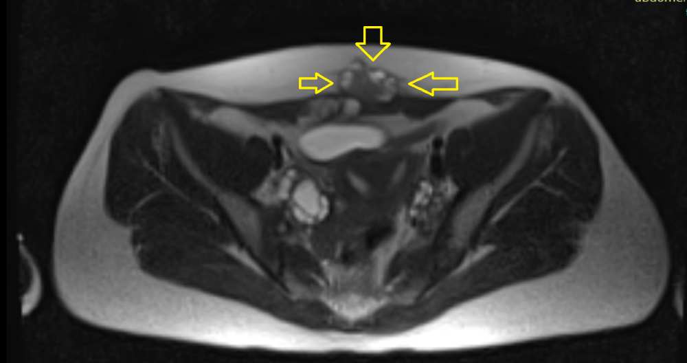
Medical history (a previous caesarean section or other operation, pain and/or a palpable mass that gets worse during the period) and clinical examination raise the suspicion. An ultrasound scan of the abdominal wall is usually the first-line test: the lesion appears as a solid nodule with irregular borders, and pressing on it with the ultrasound probe typically causes pain7. An MRI scan helps assess how deep the lesion extends (for example, whether it infiltrates the muscles or the fascia), which is very important for planning surgery.
Equally important: abdominal wall endometriosis may co-exist with pelvic endometriosis, but not always. In some patient series, this applies to about one in three women, particularly when the lesion is in the umbilicus and there has been no previous surgery8. For this reason, the work-up should also include a specialist transvaginal ultrasound scan to rule out or diagnose pelvic endometriosis.
How is it treated?
Medical treatment. Hormonal medications may relieve pain temporarily, but they do not make the lesion disappear, and symptoms usually return once they are stopped. Evidence on their long-term effectiveness is limited4.
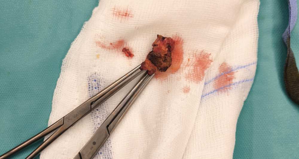
Surgical excision. This is the treatment of choice. The lesion is removed completely, with a margin of healthy tissue around it. With complete excision, the cure rate exceeds 95% and the risk of recurrence is low2. When endometriosis infiltrates the fascia of the abdominal muscles and/or the abdominal muscles themselves, a mesh may be needed to repair the abdominal wall and reduce the risk of a postoperative hernia. This is usually needed in a small proportion of patients6. In every case, accurate preoperative imaging and planning are the key to complete excision.
Newer, non-surgical methods. In recent years, techniques such as percutaneous cryoablation (freezing the lesion through a needle under imaging guidance) and high-intensity focused ultrasound (HIFU) have been studied. Early results are encouraging, but they come from small, retrospective studies, and more data are needed before these methods can be considered equivalent to surgery9.
Is there a risk of cancer?
Malignant transformation of abdominal wall endometriosis is extremely rare. In a systematic review, the total number of cases described in the international literature did not exceed 5010. However, a nodule in a scar that grows quickly, especially many years after the operation, requires urgent investigation and removal.
Can it be prevented?
Simple measures have been proposed during a caesarean section or other operations, such as protecting and cleaning the wound before closure and using clean instruments to close the abdominal wall. Their effectiveness, however, has not been proven in large studies3.
When should you be concerned?
See a gynaecologist with experience in endometriosis if you notice:
- a painful lump near or on the scar of a caesarean section or other operation,
- pain or swelling in the scar that gets worse during your period,
- a nodule in the umbilicus that hurts or bleeds during your period,
- swelling in the groin that changes with your cycle.
If you have endometriosis or any of the symptoms above, do not hesitate to contact us. Led by endometriosis gynaecologist-surgeon Dr Georgios Grigoriadis, we have a fully trained team of healthcare professionals, ready to treat endometriosis in any location, from the simplest to the most complex.
Endometriosis is common… get informed!
References
- Gruber TM, Lange K, Ebeling GS, Henrich W, Mechsner S. Scar endometriosis, a form of abdominal wall endometriosis – a neglected obstetrical complication? Arch Gynecol Obstet. 2024. doi:10.1007/s00404-024-07834-2
- Horton JD, DeZee KJ, Ahnfeldt EP, Wagner M. Abdominal wall endometriosis: a surgeon’s perspective and review of 445 cases. Am J Surg. 2008;196(2):207-212.
- Zhang P, Sun Y, Zhang C, et al. Cesarean scar endometriosis: presentation of 198 cases and literature review. BMC Womens Health. 2019;19:14. doi:10.1186/s12905-019-0711-8
- Dridi D, et al. Umbilical endometriosis: a systematic literature review and pathogenic theory proposal. J Clin Med. 2022;11(4):995. doi:10.3390/jcm11040995
- Dalkalitsis A, Salta S, Tsakiridis I, et al. Inguinal endometriosis: a systematic review. Taiwan J Obstet Gynecol. 2022;61(1):24-33.
- Ecker AM, Donnellan NM, Shepherd JP, Lee TT. Abdominal wall endometriosis: 12 years of experience at a large academic institution. Am J Obstet Gynecol. 2014;211(4):363.e1-5.
- Savelli L, Manuzzi L, Di Donato N, et al. Endometriosis of the abdominal wall: ultrasonographic and Doppler characteristics. Ultrasound Obstet Gynecol. 2012;39(3):336-340.
- Marras S, Pluchino N, Petignat P, et al. Abdominal wall endometriosis: an 11-year retrospective observational cohort study. Eur J Obstet Gynecol Reprod Biol X. 2019;4:100096.
- Bodard S, Razakamanantsoa L, Geevarghese R, et al. Percutaneous cryoablation of abdominal wall endometriosis: a systematic literature review of safety and efficacy. Insights Imaging. 2024;15:282.
- Liu G, Ren F, et al. Malignant transformation of abdominal wall endometriosis: a systematic review of the epidemiology, diagnosis, treatment, and outcomes. Eur J Obstet Gynecol Reprod Biol. 2021. doi:10.1016/j.ejogrb.2021.08.006